1. Parts of Pituitary Gland

Posterior pituitary - neurohypophysis
The hypothalamus is connected to the pit gland via pit stalk aka infundibulum.
2. Relations of the pit gland
Anterior, and superior of the gland -> optic chiasma (where 2 optic nerves of the temporal half of both eyes cross over each other and enter the optic tract.
** When pit gland enlarges, the optic chiasma will be compressed, and affect the temporal half of the vision of both eyes and result in bitemporal hemianopia.
Anterior to pituitary gland -> sphenoidal air sinus
** pit gland tumour can rupture the roof of the sphenoidal air sinus.

■■ Anteriorly: The sphenoid sinus (Fig. 11.13)
■■ Posteriorly: The dorsum sellae, the basilar artery, and the pons
■■ Superiorly: The diaphragma sellae, which has a central aperture that allows the passage of the infundibulum. The diaphragma sellae separates the anterior lobe from the optic chiasma (Fig. 11.108).
■■ Inferiorly: The body of the sphenoid, with its sphenoid air sinuses
Oculomotor nerve (CV III)
Trochlear nerve (CV IV)
Ophthalmic (VI) & maxillary nerve (VII) (CV V)
Abducens nerve (CV VI)
**When the pituitary gland enlarges, the oculomotor nerve will be compressed.
3. Development of the pit gland
The pituitary gland develops from a small ectodermal diverticulum (Rathke’s pouch), which grows superiorly from
the roof of the stomodeum immediately anterior to the buccopharyngeal membrane and a small ectodermal diverticulum (the
infundibulum), which grows inferiorly from the floor of the diencephalon of the brain.
During the second month of development, Rathke’s pouch comes into contact with the anterior surface of the infundibulum, and its connection with the oral epithelium elongates, narrows, and finally disappears Rathke’s pouch now is a vesicle that flattens itself around the anterior and lateral suraces of the infundibulum. The cells of the anterior wall of the vesicle proliferate and form the pars anterior of the pituitary; from the vesicle’s upper part, there is a cellular extension that grows superiorly and around the stalk of the infundibulum, forming the pars tuberalis. The cells of the posterior wall of the vesicle never develop extensively; they form the pars intermedia. Some of the cells later migrate anteriorly into the pars anterior. The cavity of the vesicle is reduced to a narrow cleft, which may disappear completely. Meanwhile, the infundibulum has differentiated into the stalk and pars nervosa of the pituitary gland.
4. Microscopic structure of the pit gland
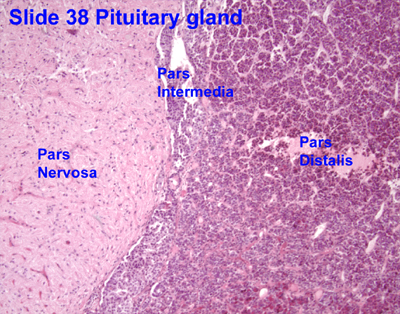
Neurohypophysis
Pars Nervosa


- axons terminate in median eminence @primary capillary plexus
- superior hypophyseal artery also meets at the primary capillary plexus
- portal veins carries hormones from primary to secondary capillary plexus
- hormones secreted by adenohypophysis goes into general circulation

8. Pit Dwarfism, gigantism, acromegaly
During the second month of development, Rathke’s pouch comes into contact with the anterior surface of the infundibulum, and its connection with the oral epithelium elongates, narrows, and finally disappears Rathke’s pouch now is a vesicle that flattens itself around the anterior and lateral suraces of the infundibulum. The cells of the anterior wall of the vesicle proliferate and form the pars anterior of the pituitary; from the vesicle’s upper part, there is a cellular extension that grows superiorly and around the stalk of the infundibulum, forming the pars tuberalis. The cells of the posterior wall of the vesicle never develop extensively; they form the pars intermedia. Some of the cells later migrate anteriorly into the pars anterior. The cavity of the vesicle is reduced to a narrow cleft, which may disappear completely. Meanwhile, the infundibulum has differentiated into the stalk and pars nervosa of the pituitary gland.
4. Microscopic structure of the pit gland
Adenohypophysis with Pars Distalis
- chromophobe cells
- chromophil cells
- Acidophils (alpha cells)
- red staining (eosin) granules in the cytoplasm and blue nuclei
- are somatrophs and mammotrophs
- As somatrophs, they secrete somatotropin, aka GH
- As mammotrophs, they secrete prolactin
- Basophils (beta cells)
- less numerous
- blue staining (heamtoxylin) granules in cytoplasm
- are thyrotrophs (TSH) , gonadotrophs (LH and FSH) and corticotrophs (ACTH)
Pars intermedia
- colloid filled follicles
- basophils- Melanocyte stimulating hormone
Neurohypophysis
Pars Nervosa
- nerve fibres
- pituicytes (unmyelinated axons and supportive cells)
- axons from neurons of (both in hypothalamus)
- paraventricular nucleus - produce oxytocin - milk ejection + contraction of uterus smooth muscle during parturition
- supraoptic nucleus - anti diuretic hormone - increases permeability of CT to water
- Herring Bodies: storage sites of the neurosecretory material of the pars nervosa neurons- contain many greyish-brown storage vesicles.
- No hormone producing cells
- Unmyelinated axons- hypothalamohypophysial tract
Pars tuberalis- surrounds the stalk, highly cellular
5. Hormones secreted by the pit gland
- Anterior- adenohypophysis
- GH
- ACTH
- Prolactin
- LH and FSH
- TSH
- Posterior- neurohypophysis
- Oxytocin
- ADH
6. How does hypothalamus regulate the secretion of hormones from pit gland?
The secretion of tropic hormones from the pituitary gland is regulated by releasing hormones from the hypothalamus, which either stimulate or inhibit the anterior pituitary gland.
7A. Hypothalamo-hypophyseal system
Hormones (Oxytocin and ADH) are produced at the paraventricular and supraoptic nuclei. They are then transported down the axons and accumulate at Herring bodies. Herring bodies has many neurosecretory granules. When released, they enter the fenestrated capillaries and pars nervosa.
7B. Hypophysial portal circulation
- superior hypophyseal artery also meets at the primary capillary plexus
- portal veins carries hormones from primary to secondary capillary plexus
- hormones secreted by adenohypophysis goes into general circulation
8. Pit Dwarfism, gigantism, acromegaly
- Pit dwarfism: deficient in GH- short
- Gigantism: excess GH- tall (child)
- Acromegaly: excess GH (adult)
9. Consequences of pit tumour
- can compress optic chiasma (located superior and anterior to pit gland) and result in bitemporal hemianopia
- can compress cavernous sinus and result in paralysis of eye muscle
10. Diabetes Insipidus (Neurogenic)
Damage to hypothalamic neuron that produces ADH- decreased amounts of ADH produced -> lots of urine secreted
Thank you! Those images of the hypothalamic nuclei responsible for the release of trophic hormones were useful.
ReplyDelete