Sunday 8 November 2015

Thyroid gland

1. Where is the thyroid gland?
In front of the neck (in front of the trachea and extends up to the thyroid cartilage)

2. Parts of the thyroid gland

True capsule- connective tissue capsule
False capsule- attached to the thyroid cartilage; gland moves up while swallowing
* Upper end of the pretracheal fascia is attached to the oblique thyroid cartilage, and connected to larynx so it moves up while swallowing

Relations

Anterior: strap muscles
Posterior: trachea and esophagus
Posterolateral: carotid arteries and internal jugular vein
Between tracheoesophageal groove: RLN

Retracting/ cutting sternohyoid and sternothyroid muscles will expose the thyroid gland

Parathyroid glands (2 pairs)

- mostly @ posterior surface of the thyroid gland/ sometimes embedded in gland
- Superior pair from 4th branchial arch
- Inferior pair from 3rd branchial arch
- Has chief cells (dark staining, arranged in clumps/ cords; secrete PTH) and oxyphil cells (scanty, pale staining)


3. Arterial supply of thyroid gland

  • superior thyroid artery from external carotid artery
  • inferior thyroid artery from internal carotid artery


Recurrent laryngeal nerve passes in between the branches of the inferior thyroid artery
  • can be damaged during thyroid surgery
  • if one side damaged: husky voice
  • if both sides: no voice and stridor


4. Venous drainage of the thyroid gland


  • Superior thyroid vein - internal jugular vein
  • Middle thyroid vein- internal jugular vein
  • inferior jugular vein - brachiocephalic vein

5. lymphatic drainage of the thyroid gland

Papillary carcinoma of the thyroid glands will spread to the lymph nodes

6. Microscopic structure of the thyroid gland
  • Arranged as follicles lined by cuboidal epithelium
  • follicular cells secrete thyroxine
  • thyroxin binds to globulin = thryroglobulin aka colloid
  • Parafollicular cells- secrete calcitonin to reduce plasma calcium levels
    • medullary carcinoma


7. Development of the thyroid gland

Physiologically, the thyroglossal duct will close, but if not -> thyroglossal fistula
If parts of the tract persist -> thyroglossal cysts
If thyroid tissue remains along the tract -> ectopic thyroid

Diseases & Evaluation of the HPT Axis

1a. Pathophysiological basis and effects of hyper and hypo secretion states of the pit hormones
POSTERIOR PITUITARY: ADH aka Vasopressin

  • ADH Excess- SIADH (lots of water is reabsorbed into plasma hence urine vol is low and concn is high)
    • concentrated urine = inappropriately high urine osmalality
    • diluted plasma = low plasma osmolality (<250 mOsm/ kg)
      • leads to water intoxification= headache, vomiting, coma, confusion due to increased intracranial pressure
      • leads to hyponatremia
    • volume expansion due to increased ECF volume of more than 10%
      • decreased aldosterone secretion
      • Increased sodium secretion (salt wasting)
        • dilutional hyponatremia -> absolute hyponatremia
    • Diagnosis of SIADH:
      • Inappropriate high urine osm (blood sample very concentrated)
      • Low plasma osm
      • hyponatremia
      • No need to measure serum AVP
    • Management
      • Restrict water intake
      • Block renal action of AVP by administering
        • demeclocycline: interferes with cAMP production
        • V2 receptor antagonists: acts on CDs
        • Replace sodium lost through urine
        • treat the cause of SIADH
          • Ectopic production of AVP by carcinoma of the lungs, pancreas
          • CNS: trauma, infections, tumours
          • Drug induced: chlopropamide, nicotine, morphine
  • Deficiency- DI (very little water is reabsorbed into plasma hence urine vol is high and concn is low; plasma volume is very low = high osmolality)
    • Cranial (Central/ Neurogenic) DI: defective/ decreased production of AVP by the hypothalamus-neuropophyseal system (hypothalamus/ pituitary)
    • Nephrogenic DI: 
      • defective renal response to AVP (hormone resistance)
        • defective V2 AVP receptors in the kidneys (receptor disease)
        • defective water channel proteins (aquaporins)


SIADH
- retention of water in excess of solutes in the body

DI
- Water diuresis -> polyuria
- Increase plasma osmolality and decrease plasma volume -> Thirst centre in the hypothalamus and leads to thirst and polydipsia

2. Primary, secondary and tertiary dysfunction
Endocrinopathies are classified as primary, secondary, or tertiary.

  1. Primary endocrine disease inhibits the action of downstream glands/ target endocrine glands. 
  2. Secondary endocrine disease is indicative of a problem with the pituitary gland. 
  3. Tertiary endocrine disease is associated with dysfunction of the hypothalamus and its releasing hormones.


3. Principles underlying the basal and dynamic tests for Hypothalamus-Hypophysis-target endocrine gland axis dysfunction
4. Principles underlying stimulation and suppression tests

Dynamic tests for AVP

  • Raise plasma osm and monitor with
    • plasma AVP secretion increases
  • To increase plasma osm
    • water deprivation
    • infusion of hypertonic saline
Stimulus for AVP secretion
- increased plasma osm
- decreased plasma volume and decreased BP


Water deprivation test to confirm if it's cranial or nephrogenic DI
  • Conducted for 8 hours during the day
  • serum and urine osm and body weight recorded every 2 hours up to 8 hours
  • fluid is not given during the test
  • DI diagnosed if
    • serum osm rises to >305mOsm/ kg (normal is from 275-295) AND
    • urine osm remains low <300 mOSM/ kg - failure of antidiuresis
  • To differentiate between cranial and nephronegic DI
    • administer synthetic AVP analogue called DDAVP through IM (des-amino-des-aspartate AVP aka desmopressin) which has more renal effect than vasoconstrictor effect
      • If urine now becomes concentrated due to actions of DDAVP on distal nephron -> Cranial DI
      • If urine still fails to concentrate as distal nephron is unresponsive to DDAVP -> Nephrogenic DI


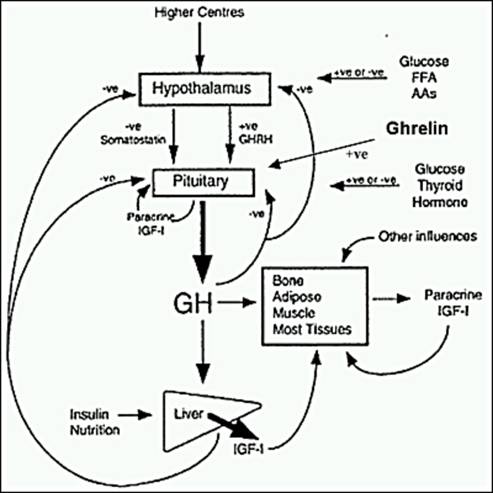
1b. Pathophysiological basis and effects of hyper and hypo secretion states of the pit hormones
ANTERIOR PIT: GH and Prolactin

Dwarfism

    • Due to GH deficiency
    • Present with systemic illness
    • deprivation/ psychosocial dwarfism: mental retardation
    • late/ no pubertal development
    • low IQ
    • unproportionate body
  • Laron dwarfs
    • hGH receptor defect in liver leading to decreased IGF-1 release leading to decreased growths
    • GH levels are normal
    • But they are not as short as pituitary dwarfs
  • Lab tests
    • measure serum GH levels after stimulating the growth hormone secretion -> dynamic provocative (stimulation) tests
      • Insulin tolerance test (gold standard)- induce hypoglycaemia (metabolic stress) - under normal response, blood glucose will fall and stimulates secretion of serum GH; also when blood glucose falls, it stimulates increase in serum ACTH and cortisol levels HENCE ITT is also used to evaluate hypothalamus-hypophyseal-adrenocortical axis
      • Post exercise blood sample - induce physical and mental stress -> release GH
      • GHRH stimulation test
      • Test for GH levels after 1 hour going to sleep
      • Glucagon - less reliable than ITT
      • Arginine
      • Clonidine
    • If serum levels of GH increased -> enough reserve
    • If not -> not enough reserve -> deficiency of GH


Gigantism (child)/ Acromegaly (adult)

    • ** patient with GH secreting adenoma that started before puberty and presents after epiphyseal closure will have features of gigantism and acromegaly
    • Due to hypersecretion of GH
    • Signs and symptoms

    • Lab tests
      • Raised serum GH and IGF-1
      • Absence of nocturnal rise of GH (GH levels raised all the time)
      • Suppression test: Oral Glucose Tolerance Test
        • Normal: hyperglycaemia will suppress GH 
        • Failure to suppress GH levels -> acromegaly
      • Neuroradiological assessment of the effect of pit tumour
Hyperprolactinaemia

  • Functions of prolactin
    • Promotes lactation
      • prolactin causes breast development during pregnancy, together with other hormones eg oestrogen
      • prolactin causes milk formation and secretion from the breast (prolactin increase action of mRNA -> promotes synthesis of milk proteins eg casein and lactalbumin)
    • Antigonadotrophic effect
      • inihibits effects of gonadotropins, leading to 
        • lactational amenorrhea
        • birth spacing
    • in males: impotence
    • Clinical signs and symptoms
      • galactorrhea
      • hypogonadism -> amenorrhea, infertility
    • Lab tests
      • basal plasma prolactin levels are raised (but must exclude stress, pregnancy and drugs)
      • dynamic tests are not useful in Ddx
    • Neuroradiological assessment of the effect of pit tumour
5. Evaluation of Hypothalamus-Hypophysis-target endocrine gland axis activity (clinical, lab-hormone assays, imaging, special investigations

Hormones regulate their plasma concentration through negative feedback along the axis
  • short loop (hormones released by ant. pituitary, eg TSH inhibits hypothalamus to release TRH)
  • long loop (hormones released by target endocrine glands eg T4 or T3, inhibits ant pituitary and hypothalamus to secrete TRH and TSH
Hypothyroidism
Primary defect: T3 & T4 low, TSH and TRH normal
Secondary defect: T3 & T4 low, TSH low and TRH normal
Tertiary defect: all low

Eg defective thyroid gland - decreased T4, increased TSH and TRH


To check if it's hypothalamic or pituitary defect, use TRH stimulation test by giving injections of TRH and measure serum TSH
  • If serum TSH increases - pituitary is normal -> hypothalamic defect
  • If serum TSH doesn't increase -> pituitary defect


Congenital Hypothyroidism -> Cretinism 
  • normally due to maternal iodine deficiency
  • Large tongue
  • Coarse features
  • Mental retardation
  • Large anterior fontanelle
  • Small stature
  • Decreased activity
  • Umbilical hernia
  • Mottled, cool, dry skin
Treatment
Administer serum TSH
  • if thyroid gland is normal -> will secrete T4
  • but if thyroid gland is not -> less T4 or no T4 is secreted
    • Require Thyroid Hormone replacement therapy

Hyperthyroidism


Primary hyperthyroidism : increased T4 and T3 but decreased TSH and TRH
Secondary/ Tertiary hyperthyroidism: increased T4 and T3, increased TSH but decreased TRH
Secondary/ Tertiary hyperthyroidism: All increased

T3 suppression test
- Large dose of T3 administered 
- Normal: Negative feedback to pituitary and hypothalamus to reduce secretion of TRH and TSH
- If fails to suppress TSH secretion -> functional tumours


Panhypopituitarism
The sequence of loss of hormonal secretion:
1. hGH
2. LH (secondary hypogonadism)
3. FSH
4. ACTH (secondary adrenal insufficiency)
5. TSH (secondary hypothyroidism)

Causes
1. Genetics
2. Nine Is (Infarction (Sheehan's syndrome), Invasive, Infiltrative, Injury, Immunologic, Iatrogenic, Infectious, Idiopathic and Isolated)

Investigations
- Stimulations tests for
  • hGH (GHRH test, ITT)
  • LH, FSH (GnRH test)
  • ACTH (CRH test, ITT)
  • TSH (free T3, T4/ TSH/ TRH)
  • Or the combination of the above: sequential injections of GHRH, CRH, GnRH and then measure serum GH< ACTH, LH and FSH


Saturday 31 October 2015

Growth hormones

1. Synthesis of GH

  • GH aka somatotrophin
  • produced by somatotropes (adenohypophysis)
  • plasma half life 20-50mins

2. Structure of GH

  • single polypeptide hormone
  • 191 aa
  • 2 disulphide linkages
  • MW of 22kDa
  • water soluble
  • GH, prolactin, chorionic somatomammotropin have similar sequence homology

3. GH receptor

  • MW of 70kDa
  • GH binds to a cell surface receptor -> dimerisation of 2 GH receptors -> forms a dimeric complex -> activates tyrosine kinase and phosphrylation of the receptor and protein kinase on tyrosine residues
4. Regulation of GH secretion
GHRH from hypothalamus and GHIH from hypothalamus and D cells of pancreas.


5. Physiological and biochemical actions of GH

  • Growth
    • Direct effects are the result of growth hormone binding its receptor on target cells. Fat cells (adipocytes), for example, have growth hormone receptors, and growth hormone stimulates them to break down triglyceride and supresses their ability to take up and accumulate circulating lipids.

    • INDIRECT: mediated by IGF
      • stimulates cartilage grwoth
      • stimulates linear bone growth by its action on the epiphyseal growth plates of long bones
      • Width of bone also increases

  • Normal carbs, lipid, protein and mineral metabolism
    • Protein synthesis: GH increases the transport of AA into muscle cells and increase protein synthesis; increases synthesis of RNA and DNA in some tissue; resemble effects of insulin
    • Carb metabolism: antagonises effect of insulin;decreased tissue glucose uptake and decreased rate of glycolysis; increased glycogen synthesis in the liver; increases hepatic glucose production via gluconeogenesis; prolonged administration can lead to dm
    • Lipid metabolism: promotes lipolysis- increases release of FFA and glycerol from adipose tissue; increases circulating FFA, causes increased oxidation of FFA in liver (ketogenic): encourages use of fat as fuel and conserves glucose
    • energy metabolism: GH increases the availability of fatty acids, which are oxidised as energy (spares glucose and proteins)
    • Its effects on tissue/ organs
      • adipose tissue: GH stimulates lipolysis -> breakdown of TG releases FFA and glycerol into the blood-> reduced synthesis of TG in fat cells
      • muscle: GH stimulates lipolysis -> increase FFA in blood surrounding muscle and hence they will be used as fuels, conserving glucose and spares proteins -> since glucose uptake is reduced, rate of glycolysis is reduced; GH increases transport of AA into muscle cells and increase protein synthesis
      • liver: stimulate production and release of IGF; GH increases oxidation of FA to acetyl CoA, enhancing ketogenesis; increased glycerol reaching the liver from lipolysis -> gluconeogenesis; increase glycogen synthesis in liver; glycolytic pathways are suppressed
    • Mineral metabolism: promotes positive calcium, magnesium and phosphate balance; retention of Na+, K+ and Cl- 
    • As it binds to lactogenic receptors, it can stimulate mammary gland eg lactogenesis (prolactin like effects)
5. Abnormal secretion of GH

  • Hyposecretion
    • GH deficient dwarfs       : Low GH and responds to exogenous GH
    • Pygmies                          : Normal GH, but low IGF-1; post GH receptor defect
    • Laron type dwarfs          : High GH but low IGF-1 and IGF-2; lack functional hepatic GH receptor
    • fail to increase GH levels in response to hypoglycaemia
  • Hypersecretion
    • most often due to pituitary tumour 
    • Children- gigantism - before epiphyseal plates close
    • Adults- acromegaly - acral bone growth causes protruded jaw, enlarged nose, hands, feet and skull
    • fail to suppress GH levels in response to glucose administration

Friday 30 October 2015

Pituitary tumours

1. Different types of Pit Tumours + its clinical features

  • Pit adenomas
    • most common
    • peak incidence 35-60yo
    • 3 types
      • Functioning: hormone excess and cause clinical manifestation 
      • Non functioning aka silent: demo of hormone production at tissue level only, without clinical manifestations of hormone excess- may cause hypopituitarism as it encroach and destroy adjacent anterior it parenchyma
      • (Both functioning and non functioning pit adenomas are composed a single cell type and produce a single predominant hormone but there are exceptions eg mammosomatotroph- excess GH and prolactin produced)
      • Hormone negative: absence of IHC reactivity/ ultrastructural evidence of hormone production 
      • (Non functioning and hormone neg adenomas are likely to present at a later stage and hence more likely to be macroadenoma.
    • incidentally diagnosed as microadenomas 
    • Classification 
      • by size (micro, macro and giant adenoma)
      • by patho 
        • ACTH cell
        • GH cell
        • prolactin cell
        • mammosomatotroph
        • TSH cell
        • Gonadotroph cell adenoma)
  • Pit carcinoma
  • Others (mets, epithelial, neural mesenchymal)

Signs and symptoms
  • Excess hormones & Mass effect
  • **Mass effect: the effect of a growing mass that results in secondary pathological effects by pushing on or displacing surrounding tissue.
    • Intracranial mass: headache and vomitting
    • hypopituitarism: compression leading to loss of normal anterior pit hormone production
    • bitemporal hermianopia
    • hyperprolactinemia due to stalk effect

Morphology
  • soft, well circumcised lesion
  • larger lesions extend and often compress the optic chiasm and adjacent structures- invasive adenomas
  • In larger adenomas, foci of haemorrhage and/ or necrosis are common
  • Histology
    • monomorphic
    • uniform round cells
    • delicate stippled chromatin -> salted pepper apperance
    • mitotic activity- scanty
Types of pit tumours
  • Prolactinoma
    • most frequent 
    • cytoplasm weakly acidophillic or chromophobe
    • sparsely/ densely granulated
    • Rupture of cells and intracellular Ca2+ leaks out and Ca2+ accumulates, resulting in dystrophic calcification -> pituitary stone
    • this tumour is characterised by 
      • efficiency (eventhough tumour is small, it will secrete hormones)
      • proportionality (size of tumour affects the amount of hormone prod)
    • Clinical signs and symptoms
      • amenorrhea
      • galactorrhea
      • loss of libido
      • infertility
      • ** Pregnancy, high dose estrogen therapy, renal failure, hypothyroidism, hypothalamic lesions and dopamine inhibiting drugs or mass in the suprasellar compartment can disturb the normal inhibitory influence of hypothalamus on prolactin secretion -> results in hyperprolactetinemia (stalk effect)
  • Growth hormone-producing adenoma
    • 2nd most common type of functional pituitary adenoma 
    • quite large when clinically symptomatic because the clinical manifestation of excessive GH is subtle
    • acidophillic
    • sparesely/ densely granulated cells
    • IHC postive for GH and cytokeratin
    • Diagnosed by increased GH and IGF-1; with glucose loading, there is no suppression of GH
  • Corticotroph cell adenoma 
    • microadenomas when diagnosed
    • stain positively with PAS stains due to carboydrate content
    • densely granulated and basophilic 
    • IHC postive for ACTH
    • Present as?
      • clinically silent
      • cause hypercortisolism due to stimulatory effect of ACTH on the adrenal cortex -> Cushing syndrome
      • cause hypercortisolism due to excessive production of ACTH by the pit -> Cushing Disease
    • large, clinically agressive corticoptroph cell adenomas may develop after surgical removal of adrenal glands for treatment of Cushing syndrome, can result in Nelson syndrome 
      • No hypercortisolism as adrenal glands are absent 
      • patients present with mass effects of the pit tumour
      • also presents in hyperpigmentation due to increased secretion of MSH
  • Gonadotroph adenomas
    • difficult to recognise 
    • secrete hormones inefficiently and variably, and secreted hormones do not cause a recognisable syndrome 
    • only detected when tumours are so large that they cause neurologic signs and symptoms
  • Thyrotroph adenomas
  • Pit adenomas (very rare!) 
    • local extension beyond the sella turcica
    • distant mets
3. Medical and surgical management of pit tumours
  • Surgery
    • transphenoidal approach esp in patients with progressive mass effect eg visual loss, hyperfunction, failure of medical treatment and pituitary apoplexy
    • must remove all cells. If not, can lead to recurrence
  • Medical
    • Prolactinoma -> dopamine agonist to reduce hyperprolactinemia & tumour size
    • GH producing adenoma -> somatastation analogues but doesn't reduce tumour size
  • Gamma knife
  • Radiation
    • esp for incomplete surgical resection, recurrent tumours, those that are unfit for surgery
    • can lead to hypopituitarism, glioma or sarcoma 
Hypopituitarism
Sheehan syndrome - postpartum necrosis of the ant pit
During pregnancy, ant pit enlarges considerably as there is a physiologic increase in the size and no of prolactin secreting cells. However, this isn't accompanied by an increase in blood supply from the low-pressure portal venous system. The enlarged gland is vulnerable to ischaemic injury esp in women who xperience significant haemorrhage during parturition. 
Post pit, as it receives blood supply directly from arterial branches (superior and inferior hypophyseal arteries),  is less susceptible. 

Pituitary apoplexy
- pit undergoes haemorrhage due to increased BP

DDx to pit tumour are Suprasellar tumours:
- induce hypo/ hyper functionaing of ant pituitary
- diabetes insipidus
- 2 main types are Gliomas from chiasm (malignant) & craniopharyngiomas (benign)


4. Craniopharyngioma (malignant transformation is rare unless exposed to radiation)
  • 2 types
    • adamantinomatous 
      • stratified squamous epitheium
      • losee reticulum
      • calcification
      • chronic inflammation
      • cholestrol rich yellow fluid -> as tumours produce lots of fats
    • papillary
      • solid and papillary sheets of squamous epithelum
      • no keratin, no calcification, no cysts
      • no reticulum

Thursday 29 October 2015

Hypothalamus & Pituitary Gland

The pituitary gland (aka hypophysis) is located below the hypothalamus, in the sella turcica. It is a fossa which can be approached through the nasal cavity and the sphenoid sinus.

1. Parts of Pituitary Gland
Anterior pituitary   - adenohypophysis
Posterior pituitary  - neurohypophysis

The hypothalamus is connected to the pit gland via pit stalk aka infundibulum.

2. Relations of the pit gland
Anterior, and superior of the gland -> optic chiasma (where 2 optic nerves of the temporal half of both eyes cross over each other and enter the optic tract.

** When pit gland enlarges, the optic chiasma will be compressed, and affect the temporal half of the vision of both eyes and result in bitemporal hemianopia.

Anterior to pituitary gland -> sphenoidal air sinus
** pit gland tumour can rupture the roof of the sphenoidal air sinus.


■■ Anteriorly: The sphenoid sinus (Fig. 11.13)
■■ Posteriorly: The dorsum sellae, the basilar artery, and the pons
■■ Superiorly: The diaphragma sellae, which has a central aperture that allows the passage of the infundibulum. The diaphragma sellae separates the anterior lobe from the optic chiasma (Fig. 11.108).
■■ Inferiorly: The body of the sphenoid, with its sphenoid air sinuses
■■ Laterally: The cavernous sinus and its contents 

Oculomotor nerve (CV III)
Trochlear nerve (CV IV)
Ophthalmic (VI) & maxillary nerve (VII) (CV V)
Abducens nerve (CV VI)
**When the pituitary gland enlarges, the oculomotor nerve will be compressed.

3. Development of the pit gland

The pituitary gland develops from a small ectodermal diverticulum (Rathke’s pouch), which grows superiorly from the roof of the stomodeum immediately anterior to the buccopharyngeal membrane and a small ectodermal diverticulum (the infundibulum), which grows inferiorly from the floor of the diencephalon of the brain. 

During the second month of development, Rathke’s pouch comes into contact with the anterior surface of the infundibulum, and its connection with the oral epithelium elongates, narrows, and finally disappears Rathke’s pouch now is a vesicle that flattens itself around the anterior and lateral suraces of the infundibulum. The cells of the anterior wall of the vesicle proliferate and form the pars anterior of the pituitary; from the vesicle’s upper part, there is a cellular extension that grows superiorly and around the stalk of the infundibulum, forming the pars tuberalis. The cells of the posterior wall of the vesicle never develop extensively; they form the pars intermedia. Some of the cells later migrate anteriorly into the pars anterior. The cavity of the vesicle is reduced to a narrow cleft, which may disappear completely. Meanwhile, the infundibulum has differentiated into the stalk and pars nervosa of the pituitary gland. 

4. Microscopic structure of the pit gland

Adenohypophysis with Pars Distalis
  • chromophobe cells
  • chromophil cells
    • Acidophils (alpha cells)
      • red staining (eosin) granules in the cytoplasm and blue nuclei 
      • are somatrophs and mammotrophs
      • As somatrophs, they secrete somatotropin, aka GH
      • As mammotrophs, they secrete prolactin 
    • Basophils (beta cells)
      • less numerous
      • blue staining (heamtoxylin) granules in cytoplasm
      • are thyrotrophs (TSH) , gonadotrophs (LH and FSH) and corticotrophs (ACTH)
Pars intermedia
  • colloid filled follicles
  • basophils- Melanocyte stimulating hormone

Neurohypophysis
Pars Nervosa
  • nerve fibres
  • pituicytes (unmyelinated axons and supportive cells)
  • axons from neurons of (both in hypothalamus)
    • paraventricular nucleus - produce oxytocin - milk ejection + contraction of uterus smooth muscle during parturition
    • supraoptic nucleus - anti diuretic hormone - increases permeability of CT to water
  • Herring Bodiesstorage sites of the neurosecretory material of the pars nervosa neurons- contain many greyish-brown storage vesicles.
  • No hormone producing cells
  • Unmyelinated axons- hypothalamohypophysial tract
Pars tuberalis- surrounds the stalk, highly cellular

5. Hormones secreted by the pit gland
  • Anterior- adenohypophysis
    • GH
    • ACTH
    • Prolactin
    • LH and FSH
    • TSH
  • Posterior- neurohypophysis
    • Oxytocin
    • ADH
6. How does hypothalamus regulate the secretion of hormones from pit gland?

 The secretion of tropic hormones from the pituitary gland is regulated by releasing hormones from the hypothalamus, which either stimulate or inhibit the anterior pituitary gland.


7A. Hypothalamo-hypophyseal system
Hormones (Oxytocin and ADH) are produced at the paraventricular and supraoptic nuclei. They are then transported down the axons and accumulate at Herring bodies. Herring bodies has many neurosecretory granules. When released, they enter the fenestrated capillaries and pars nervosa.

7B. Hypophysial portal circulation


- axons terminate in median eminence @primary capillary plexus
- superior hypophyseal artery also meets at the primary capillary plexus
- portal veins carries hormones from primary to secondary capillary plexus
- hormones secreted by adenohypophysis goes into general circulation


8. Pit Dwarfism, gigantism, acromegaly

  • Pit dwarfism: deficient in GH- short
  • Gigantism: excess GH- tall (child)
  • Acromegaly: excess GH (adult)

9. Consequences of pit tumour
  1. can compress optic chiasma (located superior and anterior to pit gland) and result in bitemporal hemianopia
  2. can compress cavernous sinus and result in paralysis of eye muscle

10. Diabetes Insipidus (Neurogenic)
Damage to hypothalamic neuron that produces ADH- decreased amounts of ADH produced -> lots of urine secreted


Tuesday 13 January 2015

Nutrition II: Micronutrients

Fat Soluble Vitamins
Vit A
Vit D
Vit E
Vit K

Water soluble vitamins
Vit C
Thiamine
Riboflavin
Niacin
Pantothenic acid
Pyroxidine
Biotin
Folate
Cobalamin

Minerals
Calcium
Zinc
Selenium
Magnesium
Iodine
Iron
Sodium
Potassium

Nutrition I: Macronutrients

Nutrients

1. Roles at cellular and molecular level

Types
Carbohydrates
  • Important source of energy
  • Provide energy to muscles- carbs have a protein-sparing action (prevent protein catabolising to provide glucose when carb levels are low and hence, can preserve muscle tissue)
  • Allows protein to perform its function (development and maintenance of muscle mass)
  • Healthy function of the CNS (CNS depends on glucose)
  • Components of glycolipids, glycoprotein and nucleic acids
  • Provide fiber: 
    • Insoluble fiber increases stool weight, promoting regular elimination of waste and prevent constipation
    • Soluble fibre: food source for gut bacteria
      • Fermentation of soluble fiber results in release of short chain fatty acids and B vitamins
      • Short chain FA block cholesterol synthesis in the liver
      • Reduce cholesterol by enhancing hepatic control to bile acid
      • Reduce postprandial rise in blood glucose
      • Delay gastric emptying and increase satiation
  • Provide desirable flavor and texture in food products
Health issues
  • dental carries
  • obesity
  • CVD
  • Colorectal cancer

Protein
  • Growth and maintenance
  • Cell structure
  • Antibodies and hormone production
  • Source of energy
  • Maintenance of fluid balance
Fat
  • source of energy
  • Supply of EFA
  • cell structure- PL
  • Required for absorption of fat soluble vitamins
  • increase palatability
Deficient of macronutrient
Carbs- increase in ketone bodies production, protein-tissue wasting
Protein- protein energy malnutrition- kwashiorkor and marasmus
Fat- weight loss, can't keep warm, lack of EFA and Vit ADEK

Excessive intake of macronutrient
Carbs- too much triglycerides in blood
Protein- proteins consumed in excess is deaminated, and the resulting carb skeletons are metabolised to provide energy/ acetyl CoA for fatty acid synthesis. Excess protein is eliminated form the body as urinary nitrogen, and is accompanied by increasing urinary calcium, leading to osteoporosis, gout etc
Fat- increased cholestrol levels leading to CVD